


A pre-targeting approach for liver radioembolisation radiation therapy, theranostics, radioembolisation, pre-targeting, interventional radiology, nuclear medicine, supramolecular chemistry, pharmaceutical industry, biotech/medical companies
radiation therapy, theranostics, radioembolisation, pre-targeting, interventional radiology, nuclear medicine, supramolecular chemistry, pharmaceutical industry, biotech/medical companies
A pre-targeting approach for liver radioembolisation

Background:
Radioembolisation is a local form of radiation-therapy that is increasingly used to treat primary liver tumours and metastases untreatable via surgery or chemotherapy. Currently, radioembolisation procedures are performed in two steps: 1) a scout-step which is used to identify (lung) shunting and to optimize dose using the non-therapeutic radiotracer 99m-technecium magroaggregate, and 2) therapeutic-step using rather costly microspheres containing therapeutic radio-isotopes (90-Ytrium or 166-Holmium).
The sequential steps are performed as two discrete procedures, usually separated because of logistical reasons by a period of two weeks. While the clinical benefit of this approach has been demonstrated, its high cost and the preclusion of procedure-related toxicity to healthy tissue e.g. lung remains a challenge. Even when using a scout scan, shunting occurs in 10% and results in the displacement of a fraction of the therapeutic microspheres outside of the diseased area, leading to ineffective dose distribution and serious adverse effects such as radiation pneumonitis. Therefore a cheaper therapeutic alternative that provides higher accuracy is needed.
Technology Overview:
Firstly, LUMC researchers have used supramolecular chemistry to develop a two-step pre-targeting approach which integrates the scout- and therapeutic-steps in a single procedure. As the therapeutic component specifically targets the diagnostic component, this prevents discrepancies in accumulation. Hence, the dose prediction becomes more accurate and the shunting issue is solved. Uniquely, the chemical interactions chosen favour complex formation within the liver, meaning that the technology is less prone to toxic side-effects due to lung shunting.
Secondly, the pre-targeting concept used creates flexibility in the therapeutic radioisotopes that can be used for the procedure. This means more easily produced radioisotopes can be used to help bring down the treatment cost. Further, the flexibility in the use of radioisotopes also allows for the creation of kit-based radioembolisation formulation that can be prepared in the hospital. With that the current therapeutic window of two weeks can be shortened to one of only hours.
Thirdly, the supramolecular chemistry used in this invention could also be adapted for use in chemoembolization approaches or for the subcutaneous needle-injection based delivery of a therapeutic dose to isolated lesions. Again, in both these indications the ability to verify the accuracy of the delivery process before administering the therapeutic component is key.
Benefits:
This technology provides a more accurate and cost effective alternative to the radioembolisation procedures currently available. The technology:
Can use a wide range of (therapeutic) radioisotopes (in addition to currently used radioisotopes), as well as chemotherapeutics, creating the potential for new markets to be explored;Improves clinical logistics by allowing one day treatments. This avoids the need for the currently used complex double procedure (scout- followed by therapeutic-procedure) spread over two weeks);Allows for kit-based radioembolisation formulations to be created and with that improves the logistics of the supply chain.
Opportunity:
We are looking for partner(s) to license the technology for commercialization to and/or to fund research into refinement and clinical translation of the technology.
Please note, header image is purely illustrative.
Source: Philip Hogeboom, NL - Wikimedia Commons - CC 3.0 Unported (CC BY 3.0)
Imaging agents that specifically target peripheral nerves surgery, image guided surgery, imaging, nerves, pharmaceutical industry, biotech/medical companies
surgery, image guided surgery, imaging, nerves, pharmaceutical industry, biotech/medical companies
Imaging agents that specifically target peripheral nerves

Background:
Damage to nerves is a common side effect of surgery that can result in loss of function. Improved visualization of nerves in the operating field, for example by using florescence nerve imaging agents, would help the surgeon avoid such accidental nerve injury. Ideally, such nerve tracers need to be specific for the peripheral nervous system with little or no cross reactivity with other tissues (adipose and central nervous tissue) as this may lead to unwanted side effects. To date there are no nerve tracers in clinical application.
Technology Overview:
Research at LUMC, funded by two grants from the European Research Council, has led to the development of a peptide-based imaging agent that specifically binds to peripheral nerves. The imaging agent targets specific markers on the membrane of myelinating Schwann cells and contains a detectable fluorescent imaging label. Consequently it is suitable for use in fluorescence-guided surgery application. The imaging potential of this agent has been validated in myelin producing cells, dorsal root ganglia cultures, and in the myelin sheaths found in peripheral nerves, both ex-vivo and in vivo.
https://www.lumc.nl/org/radiologie/research/MIIGI/imilab/grants/
http://cordis.europa.eu/project/rcn/104526_en.html
Benefits:
Fluorescence guided surgery could be used to prevent accidental nerve injury and to allow surgeons to preserve nerves during complex orthopaedic, cardiologic or oncologic interventions.
In addition, the imaging agents might also be of value in:
- the assessment of neuropathies that involve degeneration of myelin (e.g. Multiple Sclerosis)
- localization of nerves during nerve repairing surgery after trauma or plastic surgery
- evaluation of nerve tissue using immunohistochemistry (ex vivo application)
- protein detection methodologies in research (e.g. western blot, ELISA)
Opportunity:
We are looking for partner(s) to license the technology to and/or to fund research into refinement and clinical translation of the technology.
Please note, header image is purely illustrative.
Source: OpenStax Anatomy and Physiology - Wikimedia Commons - 4.0 International (CC BY 4.0)
Power tool attachment to cut an oval shape in bone medical device, power tool, orthopedics, surgery, oncology
medical device, power tool, orthopedics, surgery, oncology
Power tool attachment to cut an oval shape in bone
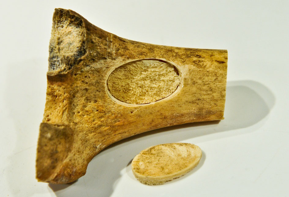
Inventors at the Leiden University Medical Center have developed a power tool attachment that cuts an oval shape.
In order to treat chondrosarcoma by curettage, an opening in bone is needed which is most often accomplished by drilling four holes and connecting them. Two disadvantages of this method are: a) the creation of sharp, squared corners which are weak points that promote breakage of the bone, and b) the loss of bone from cutting holes. This new saw creates an oval opening in the femur. This has several advantages over the conventional method. Importantly, the oval shape retains the advantage of creating a shape with a length similar to a rectangle, thus maximizing the area of the shape, but with round edges. Other advantages are that the width of the cut is 0.1 – 0.2 mm, much less than standard, thus the healing time is shortened when the bone is replaced. Furthermore less skill is needed to cut a shape in bone with an oval saw than by conventional methods.
The current length of the oval shape is approximately 3 cm but the mechanism can be developed to cut an oval shape of a different size. A smaller cutout would allow access to tumors in smaller bones.
The bone saw may also be useful to create a bone flap for cranial surgery in which the bone will be replaced. Another application may be for harvesting bone. The attachment was designed to fit an existing hand-held power tool but can be adapted. The saw can cut multiple types of materials and therefore may be useful outside the medical device arena.
Multi Mode Grasper is all-round, smart and intuitive
Multi Mode Grasper is all-round, smart and intuitive
Graspers are of great value to manipulate objects where a worksite is difficult to reach, especially during surgery. However, existing technology requires extensive training to master these skills due to the lack of forcefeedback and non-intuitive use. A TU Delft scientist has succeeded in developing a new grasper that is far more intuitive to use.
Extensive training
A major drawback of current state of the art is the lack of force-feedback that signifies the amount of force being applied by a grasper on an object. The downfall stems from the traditional rigid-linkage design that introduces friction and backlash. As a result, highly skilled surgeons must gain confidence through extensive training or continually exchange instruments to accurately perform sensitive and robust grasping maneuvers. In fact, it has been documented that experienced surgeons rely on alternative signs to interpret the level of forces being applied, for instance, by observing tissue discoloration due to a grasp. This undeniably results in expensive training and operating systems to gain the confidence and skills necessary for surgery.


Sensitive and robust grasping
A TU Delft scientist has invented a solution that resolves the drawbacks of prior state of the art. The result is a multi-mode grasper that offers superior performance through two discrete modes of operation: (1) sensitive and (2) robust grasping. The compliant tool tip design provides continuous visual force-feedback through the deformation of elastic members. Its sensitive force capabilities can be limited for optimal safety during delicate tasks. Its flexible design also improves gripping performance as it conforms more easily around objects, eliminating pinch-points and reducing slippage. Thanks to a contact-aided mechanism, the grasper does not forfeit robust grasping capabilities. By simply advancing an outer sheath, the device is capable of transferring robust forces for rigorous tasks.
Reduction of learning curve
The invention of TU Delft also includes a novel joystick design for manual control of the multi-steerable instrument tip. Whereas a conventional joystick controls only two degrees of freedom (translation forward/backward & left/right), this new joystick-design controls four degrees of freedom (translation forward/backward & left/ right + rotation forward/backward & left/right) with a simple motion of the finger or thumb. Thanks to this new design it is possible to manipulate the 3D position and orientation of the instrument tip independently of the position and orientation of the instrument shaft.
Broad range of applications
Besides surgery, this TU Delft invention has potential for many other applications. Any working enviroment that is difficult to reach and requires manipulation of objects are potential candidates. Space and deep sea mining, government defense, and agriculture are just a few examples of its future applications.
Advantages
- Simple and intuitive to use
- Reduced learning curve, less training required
- Reduced risks for patients
- 2-in-1 grasper performs all tasks, sensitive and robust
- Continuous force-feedback
- Optimal limitation of forces
- Single piece design
- Highly scalable
- No assembly required
- Simple fabrication techniques
- Easy to clean
- Broad range of applications
Flexible Grasper: Perfect Balance between Sensitivity and Force
Flexible Grasper: Perfect Balance between Sensitivity and Force
For surgeons, the ideal grasper can feel what is being grasped when performing precise and robust surgery. A grasper developed at TU Delft offers a perfect balance between sensitivity and force.
Surgeons use graspers in minimal invasive surgery, where instruments are inserted through small incisions in the abdominal wall to diagnose and manipulate tissue. Current graspers are rigid instruments that provide surgeons with very little force feedback, or sensory information about what is being grasped and the forces they are exerting. These instruments lack sensitivity between the actuation handle and the tool tip of the instrument because they consist of separate parts linked together with pinhole joints.
This design introduces backlash and friction that distorts the surgeon’s senses when gripping an organ or tumor. Due to this lack of force feedback, surgeons need extensive training before they are able to perform minimal invasive surgery. Yet, inadvertent tissue damage still occurs in surgery. Highly skilled surgeons develop alternative methods to estimate the forces they apply, for instance, by relying on discoloration of tissue. This skill can take a surgeons career to master, with little standard of consistency

Flexible
A TU Delft scientist has invented a compliant grasper that provides surgeons with a much better sense of what is being grasped. Its single-piece design eliminates friction and backlash, while simplifying fabrication, part-assembly, and sterilization. Another major improvement is the creative use of flexible members within the instrument. As a surgeon exerts force with this grasper, flexible members deform as a function of the applied force.
In this way a surgeon is able to observe the precise forces, further enhancing the feel of what he’s doing: the surgeon gains sensitivity of applied forces. This gives a surgeon better control over the forces he is exerting during precise grasping. Because of the instruments flexible design, the gripping jaws conform better to the shape of the object that is being grasped. This human-like grasping not only reduces slippage, but also further enhances usability through intuitive use.
Robust
The flexible members can be customized to the delicacy of any task. For robust forces, this grasper switches to a more powerful mode. For this purpose, the grasper features an outer sheath that can be moved forward. The tool tip contains shape-locking elements that shift into contact when the sheath is advanced.
This transmits large forces to a precisely formed grip. This feature can be used for fixating tissue, and can be quickly retracted to maintain the sensitivity of the instrument. This new grasper is also useful in other fields where objects are manipulated in difficult to reach places. In deep-sea mining, this device can be useful for distinguishing delicate artefact or hard rock. The same applies for space mining, for instance, the Mars Rover. A flexible grasper that can easily be switched from sensitive to robust modes offers possibilities in applications far beyond minimal invasive surgery
A Pre-Targeting Approach for Liver Radioembolisation A two-step pre-targeting approach for radiation therapy with increased accuracy which is less prone to toxic side-effects.
A two-step pre-targeting approach for radiation therapy with increased accuracy which is less prone to toxic side-effects.
A Pre-Targeting Approach for Liver Radioembolisation
Background
Radioembolisation is a local form of radiation-therapy that is increasingly used to treat primary liver tumours and metastases untreatable via surgery or chemotherapy. Currently, radioembolisation procedures are performed in two steps: 1) a scout-step which is used to identify (lung) shunting and to optimize dose using the non-therapeutic radiotracer 99m-technecium magroaggregate, and 2) therapeutic-step using rather costly microspheres containing therapeutic radio-isotopes (90-Ytrium or 166-Holmium).The sequential steps are performed as two discrete procedures, usually separated because of logistical reasons by a period of two weeks. While the clinical benefit of this approach has been demonstrated, its high cost and the preclusion of procedure-related toxicity to healthy tissue e.g. lung remains a challenge. Even when using a scout scan, shunting occurs in 10% and results in the displacement of a fraction of the therapeutic microspheres outside of the diseased area, leading to ineffective dose distribution and serious adverse effects such as radiation pneumonitis. Therefore a cheaper therapeutic alternative that provides higher accuracy is needed.
Technology Overview
Firstly, LUMC researchers have used supramolecular chemistry to develop a two-step pre-targeting approach which integrates the scout- and therapeutic-steps in a single procedure. As the therapeutic component specifically targets the diagnostic component, this prevents discrepancies in accumulation. Hence, the dose prediction becomes more accurate and the shunting issue is solved. Uniquely, the chemical interactions chosen favour complex formation within the liver, meaning that the technology is less prone to toxic side-effects due to lung shunting. Secondly, the pre-targeting concept used creates flexibility in the therapeutic radioisotopes that can be used for the procedure. This means more easily produced radioisotopes can be used to help bring down the treatment cost. Further, the flexibility in the use of radioisotopes also allows for the creation of kit-based radioembolisation formulation that can be prepared in the hospital. With that the current therapeutic window of two weeks can be shortened to one of only hours. Thirdly, the supramolecular chemistry used in this invention could also be adapted for use in chemoembolization approaches or for the subcutaneous needle-injection based delivery of a therapeutic dose to isolated lesions. Again, in both these indications the ability to verify the accuracy of the delivery process before administering the therapeutic component is key.
Opportunity
We are looking for partner(s) to license the technology for commercialization to and/or to fund research into refinement and clinical translation of the technology.
Keywords: radiation therapy, theranostics, radioembolisation, pre-targeting, interventional radiology, nuclear medicine, supramolecular chemistry, pharmaceutical industry, biotech/medical companies
Please note, header image is purely illustrative.
Source: Philip Hogeboom, NL - Wikimedia Commons - CC 3.0 Unported (CC BY 3.0)
Imaging Agents that Specifically Target Peripheral Nerves A peptide-based imaging agent that specifically binds to peripheral nerves, suitable for use in fluorescence-guided surgery application.
A peptide-based imaging agent that specifically binds to peripheral nerves, suitable for use in fluorescence-guided surgery application.
Imaging Agents that Specifically Target Peripheral Nerves
Background
Damage to nerves is a common side effect or surgery that can result in loss of function. Improved visualization of nerves in the operating field, for example by using florescence nerve imaging agents, would help the surgeon avoid such an accidental nerve injury. Ideally, such nerve tracers need to be specific for the peripheral nervous system with little or no cross reactivity with other tissues (adipose and central nervous tissue) as this may lead to unwanted side effects. To date there are no nerve tracers in clinical application.
Technology Overview
Research at LUMC, funded by two grants from the European Research Council, has led to the development of a peptide-based imaging agent that specifically binds to peripheral nerves. The imaging agent targets specific markers on the membrane of myelinating Schwann cells and contains a detectable fluorescent imaging label. This is suitable for use in a fluorescence-guided surgery application. The imaging potential of this agent has been validated in myelin producing cells, dorsal root ganglia cultures, and in the myelin sheaths found in peripheral nerves, both ex-vivo and in vivo.
https://www.lumc.nl/org/radiologie/research/MIIGI/imilab/grants/
http://cordis.europa.eu/project/rcn/104526_en.html
Applications
Fluorescence guided surgery could be used to prevent accidental nerve injury and allow surgeons to preserve nerves during complex orthopedic, cardiologic or oncologic interventions.
Opportunity
We are looking for partner (s) to license the technology to and / or to fund research into refinement and clinical translation of the technology.
Keywords: surgery, image guided surgery, imaging, nerves, pharmaceutical industry, biotech / medical companies
Photoactivatable Anticancer Prodrug A novel photo activated anticancer prodrug consisting in a rigidin analogue caged by a ruthenium polypyridyl complex
A novel photo activated anticancer prodrug consisting in a rigidin analogue caged by a ruthenium polypyridyl complex
Photoactivatable Anticancer Prodrug
Background
In oncology non-resectable, non-metastasized tumor treatment currently relies on chemotherapy, radiation therapy, or photodynamic therapy (PDT). Chemotherapy can be effective but it can be done because of side effects (neuron damage, pain, fatigue, etc.). Radiation therapy and PDT are more local and lower systemic toxicity but they require dioxygen in the irradiated tissues to be efficient. Hypoxic tumors, subset or tumors with high volume or hypoxic, poorly vascularized tissues, low prognosis for the patient.
The present invention is a form of photoactivated chemotherapy (PACT) that combines the advantages of well-defined target (PDT) and PDT (local activation by light and lower side effects). It is more specifically aimed at treating hypoxic tumors.
Technology Overview
The technology relies on the photochemical breakage of a chemical bond. In the prodrug form) a toxic, thioether-containing microtubule polymerization inhibitor is coordinated to a non-toxic ruthenium (II) caging complex. In the dark, the coordination bond between Ru 2+ and the sulfur atom is stable, but under light irradiation is broken, and the non-toxic caging group.
Details and State of Development:
- Collaboration between Leiden University, NL, and Texas State University, USA.
- Uncaged microtubule inhibitor patented by Prof. Alexander Kornienko from Texas State University, USA
- Synthesis, photochemistry, dark stability, and microtubule polymerization inhibition upon light irradiation, have been demonstrated
Chromatography-free synthesis available
- Low dark toxicity and high toxicity in vitro in 2D human cancer cell monolayers demonstrated both in normoxic (21% O 2 ) and hypoxic (1% O 2 ) conditions
- Low dark toxicity and high toxicity in vitro in A549 lung cancer 3D tumor spheroids
- Preliminary results in vivodemonstrate 30% tumor volume reduction under green light irradiation in A549 lung tumor xenografts in nude mice, and no toxicity in the dark, after intraperitoneal injection at 1 mg / kg and with a green light dose of 38 J / cm 2 .
Applications
Market for photoactivated chemotherapy includes brain, liver, head and neck, non-melanoma skin, and eye cancer (Market study available). Best application for tumors with a high ratio of hypoxic to normoxic volume, for which currently available therapies (PDT, radiation, surgery) do not work or are impossible (non-resectable tumors).
Opportunity
The researchers are looking for partners in the field of research, which would be extensive in vivo (mouse model) studies.
Tumor Associated Carbohydrate Antigens as Targets for Colorectal Cancer Immunotherapy The present invention relates to a novel method to identify highly specific tumor associated carbohydrate antigens (TACAs) derived from colorectal cancer (CRC) tissue.
The present invention relates to a novel method to identify highly specific tumor associated carbohydrate antigens (TACAs) derived from colorectal cancer (CRC) tissue.
Tumor Associated Carbohydrate Antigens as Targets for Colorectal Cancer Immunotherapy

Summary
The present invention relates to a novel method to identify highly specific tumor associated carbohydrate antigens (TACAs) derived from colorectal cancer (CRC) tissue. Furthermore, it is believed that the markers disclosed may be useful therapeutic targets easily accessible as expressed on cell surfaces directly accessible to therapeutics and can be carried by multiple
proteins, reflecting the overall glycosylation phenotype of the cell, providing a broader tumor targeting strategy.
There was a patent filed recently which covers the workflow of extracting, identifying and analysing these highly specific structures as well as another one that includes the specific TACA structures which were found to be solely present in CRC and not in healthy colon providing a number of promising targets for potential therapy.
Background
Colorectal cancer (CRC) is one of the leading malignancies worldwide with over 900,000 deaths in 20201. Conventional therapeutic strategies include chemotherapy, radiation therapy, and surgery. However, due to poor screening strategies and lack of symptoms in early stages, most cases are detected at an advanced stage, leading to unsuccessful treatment. Unraveling the glycome in cancer is important for the development of immunotherapies for treating solid tumors and the discovery of cancer-specific glycan structures is critical for improving how these cancers are targeted.
Technology
The present invention provides a better understanding of the variation in
O-glycosylation and its association with cell phenotypes. An in-depth
structural O-glycosylation analysis of 26 CRC cell lines was performed.
Additionally, the O-glycosylation signatures of primary cell lines as well as the primary and metastatic colorectal cancers they have been derived from were mapped and compared. Furthermore, glycosylation signatures of paired micro-dissected cancer and healthy mucosa were investigated. The released O-glycans were analysed on a sensitive nano-liquid chromatography coupled to a tandem mass spectrometer using electrospray ionization enabling powerful separation of isomeric species, as well as in-depth structural characterization of the epitopes. The major outcomes were as follows:
- Using transformative technology based on laser capture microdissection allowed for the first time to dissect the cancer cell O-glycome of CRC.
- Highly specific glycan structures were observed in cancer cells from most of the tumor tissue which were not present in healthy control tissue.
Value Proposition
The global colorectal cancer therapeutics market is poised to grow by USD 994.94 million during 2019-2023, progressing at a CAGR of almost 3% (www.businesswire.com).
Team
Prof Manfred Wuhrer currently holds the position as Head of the Center for Proteomics and Metabolomics (CPM) at the Leiden University Medical
Center in Leiden/ The Netherlands. Dr Guinivere Lageveen Kammeijer is
Senior Scientist at the CPM focusing on exploring cancer glycosylation for developing novel therapeutic and diagnostic applications. Dr Katarina
Madunic is Scientist at the CPM specialized in dissecting cancer cell
glycosylation.